The end of XIX century and the first half of the XX century are known for their outstanding discoveries that radically changed medicine. Not without reason it is said that medicine is divided into two fundamentally different stages: before the discovery of X-rays and after their discovery. Currently, X-ray radiography is an important, but not the only component of medical radiology. The use of other methods of radiation diagnostics, such as magnetic resonance imaging, radioisotope research, medical thermography and, of course, ultrasound diagnostics made it possible to put the diagnostic process on a scientific and even mathematical platform, which was unthinkable in the pre-X-ray era.
Ultrasound diagnostics in medicine has advanced much more slowly than radiology or radioisotope research. Thus, less than a year passed between the discovery of X-rays and their use in medical practice, but 70 years passed between the discovery of the piezoelectric effect and the design of a medical two-dimensional ultrasound apparatus, while the first mention of the diagnostic properties of ultrasonic waves was 18 years earlier than the discovery of X-rays! Back in 1877, an Englishman John Williams Strutt published the monograph “Theory of Sound”, which formed the basis of the science of ultrasound, and in 1880 Pierre and Jacques Curie made a discovery that triggered the development of ultrasound equipment – the “piezoelectric effect” was discovered. The sinking of the Titanic prompted Konstantin Shilovsky in 1915 to engage in echolocation and design a hydrophone, the prototype of a sonar, together with the Frenchman Paul Langevin. In 1927, the Soviet scientist S.Ya. Sokolov established the ability of ultrasound to propagate in problem metals, and an ultrasonic flaw detector was designed. In 1937, the Dussik brothers, a neurologist and engineer, tried to use echoscopy to diagnose a brain tumor, but the technique was unsuccessful and never developed. In 1949, Douglas Howry began to create the first medical ultrasound diagnostic device, and in 1950, a two-dimensional image of the internal organs of a person was obtained. In the early 1950s, Swedish scientists Inge Edler and Karl Hertz received the first one-dimensional, and in 1967 – two-dimensional ultrasound image of the heart, which was called echocardiography.
Back in 1842, Christian Andreas Doppler published a treatise in which he explained the different color of stars by the spectrum of a shift in white color as a result of the movement of the stars. However, physicist Shigeo Satomura (Japan) first applied the Doppler effect only 114 years later – in 1956. It was an atraumatic method for diagnosing heart valve defects! His work was devoted to the displacement of ultrasound frequencies, which was generated by the movements of the heart valve.
The problem of the spread of scientific knowledge in the middle of the last century was associated with the fact that scientific articles were published in the language of the authors: Russian, Japanese, German, French, English, therefore, they often had to “reinvent the wheel”, that is, scientists were working on a certain problem and, having no information about what was happening in other countries, came to the same results instead of moving forward. In this regard, in recent decades, all scientific articles are preceded by a summary in English, as the most simple and common language in the world.
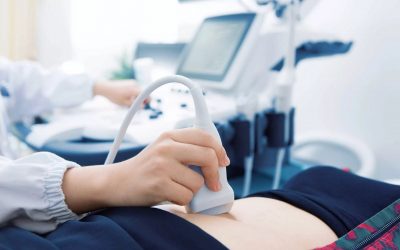
Currently, ultrasound diagnostics is the most demanded component of radiation diagnostics due to its availability, relatively inexpensive equipment and almost limitless opportunities in all areas of medical practice. New more and more advanced devices, new complex methods of ultrasound examination allow diagnosing the pathology of almost all organs and systems of a person. It should only be always remembered that the conclusion on the research carried out is not made by a machine, but by a doctor – a specialist in ultrasound diagnostics, on whose qualifications the final result depends.
Ultrasound diagnostics in medicine constantly needs young, energetic, competent specialists, and our Department of Radiation Diagnostics and Therapy of KSMU may help those who dream of becoming ultrasound doctors in obtaining basic knowledge and practical skills, and subsequently – in improving the medical art.
Stay healthy! Never stop moving forward!
Head of the Department of Radiation Diagnostics and Therapy, Doctor of Medical Sciences, Professor
Vorotyntseva Natalia Sergeevna
Resident of the Department of Radiation Diagnostics and Therapy
Petrusenko Daria Igorevna

